44 diagram and label a chromosome in prophase
Top Labeled Chromosome Images, Diagrams and Structure (Download) From the labeled chromosome diagram, you can see that a typical chromosome structure is an X-shaped complex network of DNA and protein. It has four arms; two short arms are known as "p arm" while two long arms are known as "q arms". Genes- a unit of inheritance, are located on chromosome arms. Mitosis worksheet and diagram identification.docx - Mitosis... What phase is it? prophase 15. In cell A, what is the structure labeled X? centriole 16. In cell F, what is the structure labeled Y? spindle fibers 17. Which cell is not in a phase of mitosis? d 18. What two main changes are taking place in cell B? telophase, cytokinesis 19. Sequence the six diagrams in order from first to last: d, a, f, c , e ...
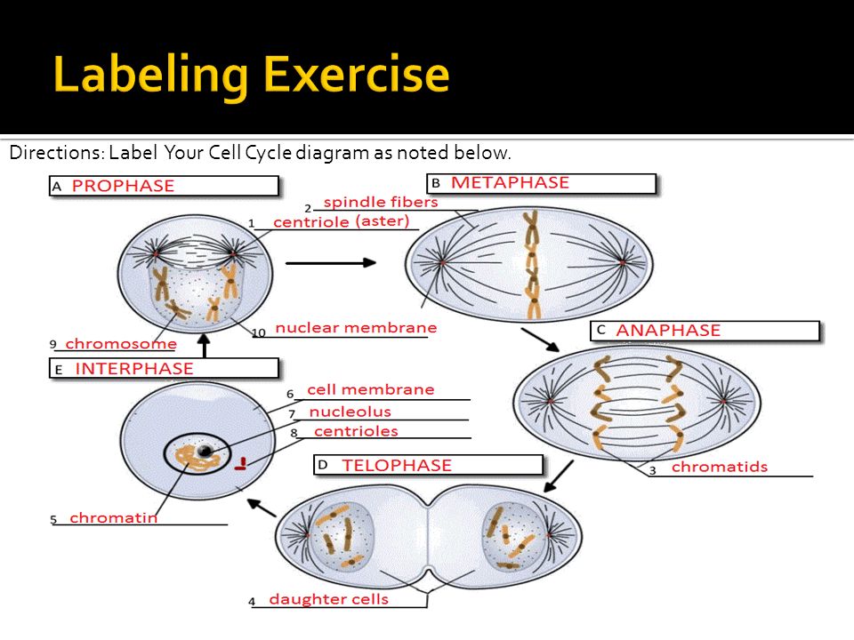
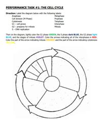
Cell Cycle Label - The Biology Corner Image shows the stages of the cell cycle, interphase, prophase, metaphase, anaphase, and telophase and asks students to name the phase and identify major structures such a centrioles and chromatids. Questions about mitosis follow the image labeling. ... If a dog cell has 72 chromosomes, how many daughter cells will be created during a single ...

Diagram and label a chromosome in prophase
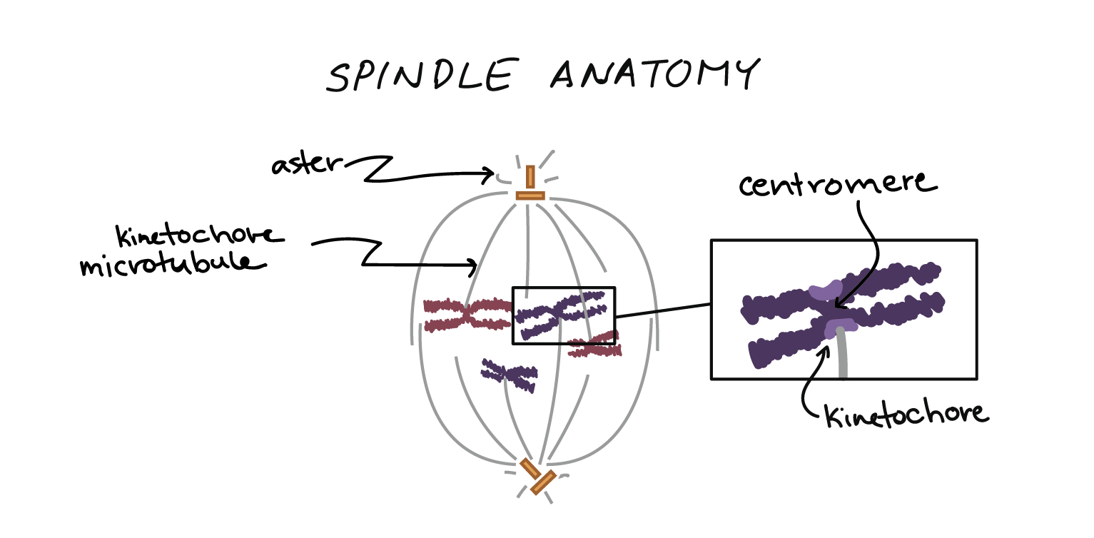
Function and Structure of Chromosomes (With Diagram) The centromere is the site of attachment of the chromosome to the microtubules of the spindle and acts as the focus of chromosome (or chromatid) movement during the anaphase of division. Chromosomes that lack a centromere are said to be acentric and fail to segregate normally during division. Secondary and tertiary constrictions may also be ... Prophase I - Definition, Stages and Quiz | Biology Dictionary Each chromosome is made up of two chromatids joined at the middle by a centromere. Each chromatid is identical.In the image below, number 1 depicts a single chromatid, 2 shows the centromere that joins both chromatids, 3 is the short (or 'p') arm and 4 the long ('q') arm of the chromosome.. Parts of a chromosome. Haploid refers to a gamete or sex cell - the spermatozoa in males and ... Prophase - Yale University Prophase Prophase At prophase the replicated chromosomes condense. Compare the staining of the chromosomes in the prophase cell to those in the surrounding, non-dividing cells. Each chromosome in prophase consists of two sister chromatids. Note that the nuclear envelope is still intact.
Diagram and label a chromosome in prophase. Prophase - Definition and Stages in Mitosis and Meiosis | Biology Prophase, in both mitosis and meiosis, is recognized by the condensing of chromosomes and separation of the centrioles in the centrosome. This organelle controls the microtubules in the cell, and each centriole is one half of the organelle. During prophase, they separate to provide microtubule centers in each new cell. Explain the structure of chromosome with diagram. - Toppr Ask The chromosome is the condensed and compactly arranged structure of the DNA with the help of histone proteins H1, H2A, H2B, H3 and H4. This is the structure which can be visible during the metaphase of cell division. This condensed packing allows the long DNA in the eukaryotes to be packed in the nucleus of the cell. Three-dimensional positioning and structure of chromosomes in a human ... We show a three-dimensional (3D) image of a human prophase nucleus obtained by serial block-face scanning electron microscopy, with 36 of the complete set of 46 chromosomes captured within it. Diagram and label a chromosome in prophase - Brainly.com Diagram and label a chromosome in prophase - Brainly.com Ii) The reason for population control is as followed1. The desire for a male child.2. Lack of awareness about birth control measures.iii) 1. The pituitary gland… heavenlute12 heavenlute12 1 week ago Biology High School answered Diagram and label a chromosome in prophase 1 See answer
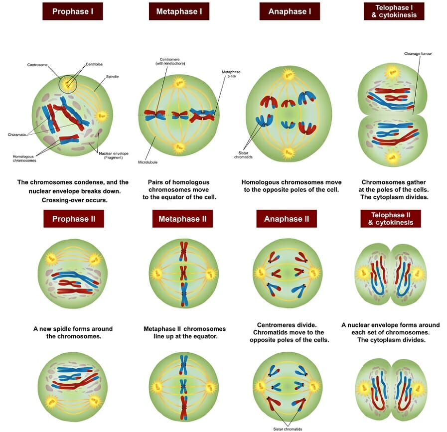
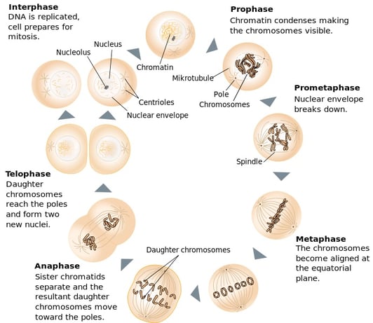
Meiosis Labeling - Google Slides They are responsible for making the spindle which will move chromosomes into position. Use the highlighted words to label the diagram, You will need to type them into the yellow boxes. The Big... The Stages of Mitosis and Cell Division - ThoughtCo In prophase, the chromatin condenses into discrete chromosomes. The nuclear envelope breaks down and spindles form at opposite poles of the cell. Prophase (versus interphase) is the first true step of the mitotic process. During prophase, a number of important changes occur: The stages of mitosis in detail - Cell division - BBC Bitesize The stages of mitosis are: prophase, metaphase, anaphase and telophase. Only two pairs of chromosomes are shown in the diagrams below. There are 23 pairs of chromosomes in a diploid human body cell. Label Chromosome Diagram | Quizlet Start studying Label Chromosome. Learn vocabulary, terms, and more with flashcards, games, and other study tools.
The fine structure of the mammalian chromosome in meiotic prophase with ... The fine structure of chromosomes in the meiotic prophase of vertebrate spermatocytes. J Biophys Biochem Cytol. 1956 Jul 25; 2 (4) ... THE ORGANIZATION AND DUPLICATION OF CHROMOSOMES AS REVEALED BY AUTORADIOGRAPHIC STUDIES USING TRITIUM-LABELED THYMIDINEE. Proc Natl Acad Sci U S A. 1957 Jan 15; 43 (1) ... Label the diagram below. Use these choices: Metaphase 1, Metaphase 2 ... First stage of Meiosis 1.The centrioles move to the poles of the cell, the nuclear membrane disintegrates, homologous chromosomes pair up (in the form of tetrad), form a chiasmata and then exchange segments of chromosomes with each other. This process is called crossing over. Metaphase 1: Solved Q5.Diagram the expected appearance of the chromosomes | Chegg.com Q5.Diagram the expected appearance of the chromosomes involved at the pachytene stage of prophase I for chromosomes 5 and 6 of the euro and red kangaroo hybrid. Label your chromosomes and include the spindles if appropriate. This question will assist you with answering Q5b. PDF Cell Division Animal Cell and Mitosis Key - Scarsdale Public Schools 1. Label the four phases of mitosis in the diagram. 2. Label the spindles and centrioles in one of the phases. 3. Color each chromosome in prophase a different color. Follow each of these chromosomes through mitosis. Show this by coloring the correct structures in each phase of mitosis. les Interphase Cytokinesis hase Chromatin
Anaphase: Definition, Checkpoints, Diagram, and Examples Anaphase: Chromosomal split forms daughter chromatids; travels to the opposite poles. The chromosomes are V - Shaped as they are dragged to the opposite sites. Telophase: Microtubules disappear and chromosomes decondense to chromatin mass. Nuclear envelope starts to form. The disintegrated organelles form again.
The 4 Mitosis Phases: Prophase, Metaphase, Anaphase, Telophase Anaphase ensures that each chromosome receives identical copies of the parent cell's DNA. The sister chromatids split apart down the middle at their centromere and become individual, identical chromosomes. Once the sister chromatids split during anaphase, they're called sister chromosomes. (They're actually more like identical twins!)
Prophase Diagrams - Wiring Diagrams Free This diagram summarises the whole process. . Stages of the Cell Cycle - Mitosis (Interphase and Prophase). by Rhys Baker. Prophase is the first stage of cell division in both mitosis and meiosis. Beginning after interphase, DNA has already been replicated when the cell enters. A summary of Prophase and Prometaphase in 's Mitosis.
A Labelled Diagram Of Meiosis with Detailed Explanation Diagram for Meiosis Meiosis is a type of cell division in which a single cell undergoes division twice to produce four haploid daughter cells. The cells produced are known as the sex cells or gametes (sperms and egg). The diagram of meiosis is beneficial for class 10 and 12 and is frequently asked in the examinations.
DOC Draw labeled diagrams showing the 5 stages of mitosis (prophase ... Draw labeled diagrams showing the 5 stages of mitosis (prophase, prometaphase, metaphase, anaphase and telophase) for a haploid cell where N=4. Use 4 different shapes to represent the different chromosomes that make up a complete set. Make sure you show the number, shape, color and location of the chromosomes at each stage of division.
Movement of Chromosomes during Anaphase (With Diagram) ADVERTISEMENTS: Read this article to learn about the Movement of Chromosomes during Anaphase ! During nuclear division or mitosis, there is a progressive change in the structure and appearance of the chromosomes. Although mitosis is a continuous process (Figs. 20-20 and 20-21), for convenience it is usually divided into four major stages: prophase, meta- phase, […]
Meiotic Division Beads Diagram How are meiosis I and meiosis II different? Configure the chromosomes as they would appear in each of the stages of meiotic division (prophase I and II, metaphase I and II, anaphase I and II, telophase I and II, and cytokinesis). Diagram the corresponding images for each stage in the section titled "Trial 2 - Meiotic Division Beads Diagram".
Meiosis I: Definition, Stages, Phases, and Diagram - Research Tweet o The chromosome is divided into "Hotspots" and "Cold spots" based on the recombination sites. o Telomeric and heterochromatin centromere regions are prevented from crossing over. o Other regions are exposed to the crossing over for the recombination of the genetic materials among the maternal and paternal genes.
Mitosis (Definition, Diagram & Stages Of Mitosis) - BYJUS The centromere of each chromosome leads at the edge while the arms trail behind it. Anaphase Telophase The chromosomes that cluster at the two poles start coalescing into an undifferentiated mass, as the nuclear envelope starts forming around it. The nucleolus, Golgi bodies and ER complex, which had disappeared after prophase start to reappear.
Prophase - Yale University Prophase Prophase At prophase the replicated chromosomes condense. Compare the staining of the chromosomes in the prophase cell to those in the surrounding, non-dividing cells. Each chromosome in prophase consists of two sister chromatids. Note that the nuclear envelope is still intact.
Prophase I - Definition, Stages and Quiz | Biology Dictionary Each chromosome is made up of two chromatids joined at the middle by a centromere. Each chromatid is identical.In the image below, number 1 depicts a single chromatid, 2 shows the centromere that joins both chromatids, 3 is the short (or 'p') arm and 4 the long ('q') arm of the chromosome.. Parts of a chromosome. Haploid refers to a gamete or sex cell - the spermatozoa in males and ...
Function and Structure of Chromosomes (With Diagram) The centromere is the site of attachment of the chromosome to the microtubules of the spindle and acts as the focus of chromosome (or chromatid) movement during the anaphase of division. Chromosomes that lack a centromere are said to be acentric and fail to segregate normally during division. Secondary and tertiary constrictions may also be ...








(89).jpg)





Post a Comment for "44 diagram and label a chromosome in prophase"