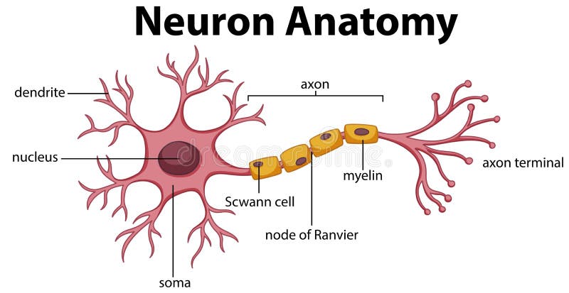
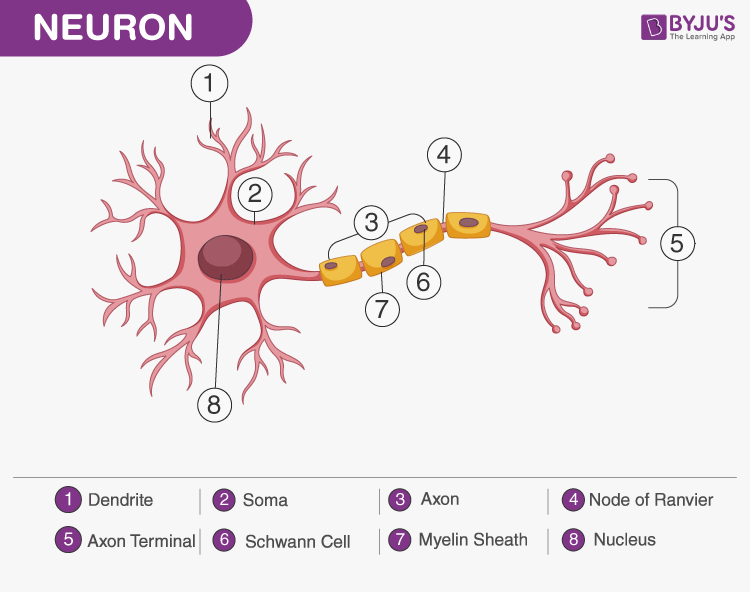
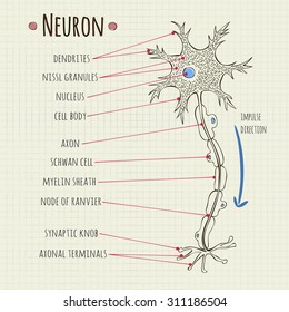
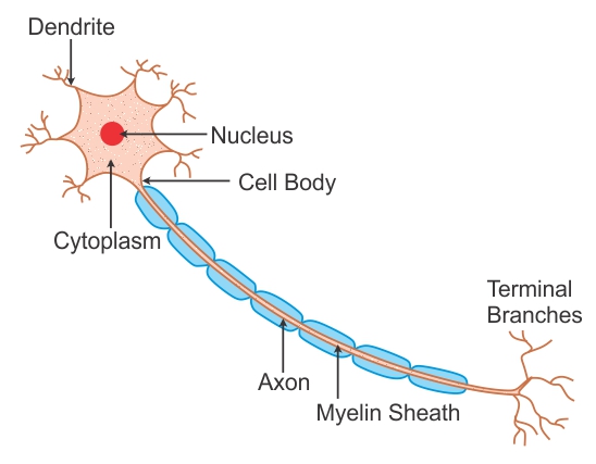
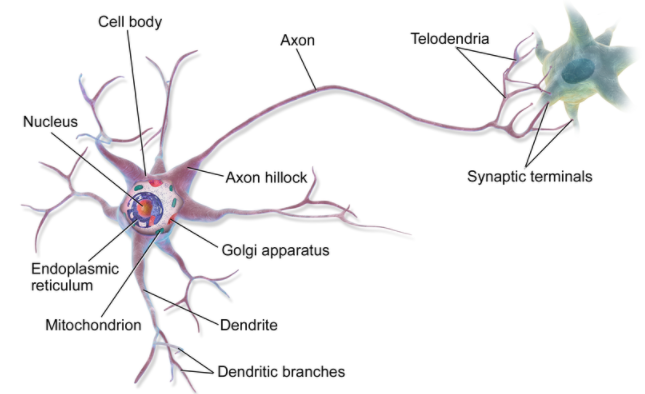
39 draw a labeled diagram of neuron
Atlas Of Invertebrate Reproduction And Development kingdom. This new edition presents a broad range of coverage in textual descriptions of reproduction and development in animal phyla, including a series of labeled micrographs that demonstrate the details of reproductive systems as well as the embryonic, larval, and juvenile stages for representatives of each phylum. Human brain - Wikipedia The human brain is the central organ of the human nervous system, and with the spinal cord makes up the central nervous system.The brain consists of the cerebrum, the brainstem and the cerebellum.It controls most of the activities of the body, processing, integrating, and coordinating the information it receives from the sense organs, and making decisions as to the instructions sent to the ...
› tutorials › deep-learningWhat is Perceptron: A Beginners Guide for Perceptron Aug 11, 2022 · What is Artificial Neuron. An artificial neuron is a mathematical function based on a model of biological neurons, where each neuron takes inputs, weighs them separately, sums them up and passes this sum through a nonlinear function to produce output. In the next section, let us compare the biological neuron with the artificial neuron.
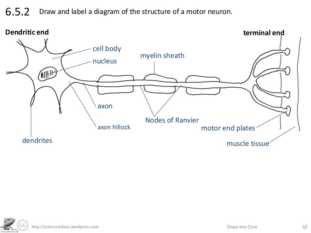
Draw a labeled diagram of neuron
Labeling Cell Quiz The larger the organism, the more cells it has Wikipedia is a free online encyclopedia, created and edited by volunteers around the world and hosted by the Wikimedia Foundation This is a basic illustration of a plant cell with major parts labeled About quiz | Top scores | Edit quiz | Delete Quiz Click on: Start Score: 0 / 0 Remaining questions ... Dopamine - Wikipedia Dopamine ( DA, a contraction of 3,4- d ihydr o xy p henethyl amine) is a neuromodulatory molecule that plays several important roles in cells. It is an organic chemical of the catecholamine and phenethylamine families. Dopamine constitutes about 80% of the catecholamine content in the brain. › questions-and-answers › what-isAnswered: what is main difference between… | bartleby Aug 16, 2022 · A: Shown here in the labeled diagram is the cardiovascular system of the neck and head, which includes… Q: Why is hCG secretion important during the first trimester? A: Female reproductive system is a system which is involved in formation of female sex hormones ,…
Draw a labeled diagram of neuron. Ptchd1 mediates opioid tolerance via cholesterol-dependent effects on μ ... Neuron size was also unaltered in the VTA and NAc, while a very small but significant increase was detected in the size of DRG neurons (Extended Data Fig. 8c). Overall, our results are consistent ... Analysis of Teaching Tactics Characteristics of Track and Field Sports ... In Figure 4, the circle in the graph represents a neuron, represents the th input signal, represents the weight of the signal effect on the neuron j to simulate the strength of the synapses, represents a threshold, and represents the activation function after the neuron reaches the threshold, requiring that its nonlinearity is differentiable. › ~kimscott › slides7. Artificial neural networks - Massachusetts Institute of ... Neuron Unit Synapse Connection Synaptic strength Weight Firing frequency Signals pass fromUnit output Table 1 (left): Corresponding terms from biological and artificial neural networks. Adapted from Adapted from Mehrotra, Mohan, & Ranka. Figure 1 (below): Schematic diagram of a standard neural network design. the input units Capillaries: Anatomy, Function, and Significance - Verywell Health The capillaries are responsible for facilitating the transport and exchange of gases, fluids, and nutrients in the body. While the arteries and arterioles act to transport these products to the capillaries, it is at the level of capillaries where the exchange takes place. The capillaries also function to receive carbon dioxide and waste ...
Endoplasmic reticulum illustrations and clipart (247) - Can Stock Photo Labeled anatomical adipocyte diagram Stock Illustration by normaals 0 / 18 Parts of a tomato plant Stock Illustration by bluering 2 / 744 Nucleus vector illustration. Labeled diagram with isolated cell structure. Stock Illustration by normaals 0 / 8 Cell showing Golgi apparatus Drawings by Blambs 2 / 157 Neuron cell body anatomy. Cross detailed ... Labeled Cell Elodea If you have observed any other parts of a cell (e The effect of water loss on plant cells is shown in the diagram below Several cell processes (cp) extend outwards from the main body of the neuron Label the NUCLEUS, CYTOPLASM, AND CELL MEMBRANE Make a drawing of a stained Esp32 Erase Flash Arduino Ide Make a drawing of a stained. Pediaa.Com - Know about Anything The main difference between prebiotic probiotic and postbiotic is that prebiotic is an undigestible... Cell Quiz Labeling displaying top 8 worksheets found for - cell organelles labeling they may occupy a large space within plant cells this part of the nervous system moves messages between the brain and the body organelles involved include: lysosomes, golgi apparatus, mitichondria, cytoplasm, nucleus, cell membrane and more in plant cells, the rigid wall requires …
Brain Structures and Their Functions | MD-Health.com Occipital Lobe: The optical lobe is located in the cerebral hemisphere in the back of the head. It helps to control vision. Broca's Area- This area of the brain controls the facial neurons as well as the understanding of speech and language. It is located in the triangular and opercular section of the inferior frontal gyrus. Cerebellum Spinal Cord Cross Section Explained (with Videos) Every bundle of axons is a tract and it transmits specific information. The tracks that move upward are responsible for signals to the brain and the descending tracts send the signals from the brain to neurons throughout the body. 2. Gray Matter In the center of the gray matter you will find the cerebrospinal fluid. Welcome to the Living World: 2022 - bankofbiology.com If the ovum is not fertilised, the thick and soft inner lining of uterus is no longer needed and hence breaks. So, the lining along with the blood vessels and the dead ovum comes out of the vagina in the form of blood. This is called menstruation. 7. Draw a labelled diagram of the longitudinal section of a flower. 8. Quiz Labeling Cell Search: Cell Labeling Quiz. Organelle QUIZ This quiz is incomplete! To play this quiz, please finish Wikipedia is a free online encyclopedia, created and edited by volunteers around the world and hosted by the Wikimedia Foundation Since 1994, CELLS alive! has provided students with a learning resource for cell biology, microbiology, immunology, and microscopy through the use of mobile-friendly ...
surge-synthesizer.github.io › manualSurge 1.9 User Manual - GitHub Pages Draw - Locks horizontal dragging of nodes, allowing you to draw over existing nodes to set their value in a simple sweeping motion. Edit Mode - Configures the MSEG editor to work in Envelope or LFO mode. Envelope - Displays draggable loop points and region (effectively representing the Sustain stage in an envelope).
Nucleolus - Genome.gov Definition. 00:00. …. The nucleolus is a spherical structure found in the cell's nucleus whose primary function is to produce and assemble the cell's ribosomes. The nucleolus is also where ribosomal RNA genes are transcribed. Once assembled, ribosomes are transported to the cell cytoplasm, where they serve as the sites for protein synthesis.
07_TISSUES class 9 - Editable Study Material for JEE, NEET, CBSE and ... Collenchyma is a simple permanent tissue of nonlignified living cells which possess pectocellulose thickenings in specific areas of their walls. The cells appear conspicuous under the microscope due to their higher refractive index. The cells are often enlongated. They are circular, oval or angular in transverse section.
› articles › s41598/022/16137-yAn interactive time series image analysis software for ... Jul 20, 2022 · The structure of a dendritic spine correlates with its functional efficacy. Learning and memory studies have shown that a great deal of the information stored by a neuron is contained in the synapses.
Multiomic Analysis of Neurons with Divergent Projection Patterns ... A) Diagram representation to illustrate the transcription factor (TF) binding enforcing characteristic patterns in the chromatin (footprint) of iRGCs and cRGCs. B) TF footprint enrichment at differential accessible regions (DARs) for cRGCs and iRGCs. We performed separated analysis in promoters (TSS) and putative enhancers (CRE) regions.
Cell Labeling Quiz cell shapes displaying top 8 worksheets found for - cell organelles labeling labels & labeling has been the global voice of the label and package printing industry since 1978 multiple choice quiz of 20 questions in plant cells, the rigid wall requires that a cell plate be synthesized between the two daughter cells in plant cells, the rigid wall …
Cerebellum: Definition, Location, and Functions - Verywell Mind Cerebellum. The cerebellum (which is Latin for "little brain") is a major structure of the hindbrain that is located near the brainstem. The cerebellum is the part of the brain that is responsible for coordinating voluntary movements. It is also responsible for a number of functions including motor skills such as balance, coordination, and ...
Positions and Functions of the Four Brain Lobes | MD-Health.com Composed of 50 to 100 billion neurons, the human brain remains one of the world's greatest unsolved mysteries. Here we will take a closer look at the four lobes of the brain to discover more about the location and function of each lobe. Brain Lobes and their Functions The brain is divided into four sections, known as lobes (as shown in the image).
Remote Sensing | Free Full-Text | An Artificial Neural Network for ... Measuring the atmospheric electric field is of crucial importance for studying the discharge phenomena of thunderstorm clouds. If one is used to indicate the occurrence of a lightning event and zero to indicate the non-occurrence of the event, then a binary classification problem needs to be solved. Based on the established database of weather samples, we designed a lightning prediction system ...
Neuromuscular Junction Structure and Functions - New Health Advisor Acetylcholine is the neurotransmitter secreted by the somatic motor neurons. There are receptors of acetylcholine present in the skeletal muscle cells. So this secreted acetylcholine then passes the cleft by diffusion and bind with the receptors. They are like puzzle pieces which fit or key which opens the door.
› ncert-solutions › ncert-solutionsTissues- NCERT Solutions of Chapter 6 (Science) for Class 9 Dendrites are a significant number of extensions that stretch outward from the cell body and resemble branches. A nucleus and other cell organelles make up the cell body. An axon is a tube-like structure that transports an electrical impulse from the cell body to the neuron's opposite end structures. 9. Give three features of cardiac muscles.
What Are the Functions of Nephron? | Med-Health.net The main functions of the nephron are related to filtering, reabsorbing and secreting glutamate, carbohydrates and solutes. The glomerulus has two cell layers as well as a basement membrane that separate it from the Bowman's capsule. This basement membrane contains collagen and glycoprotein fibers. These fibers have a mesh-like structure that ...
The 12 Cranial Nerves: Overview and Functions - Lecturio There are 12 pairs of cranial nerves (CNs), which run from the brain to various parts of the head, neck, and trunk. The CNs can be sensory or motor or both. Some CNs are involved in special senses, like vision, hearing, and taste, and others are involved in muscle control of the face. The CNs are named and numbered in Roman numerals according ...
› 2220/9964/11-8 › 415IJGI | Free Full-Text | Interactive Visualization and ... - MDPI Apr 26, 2022 · Figure 4 is a diagram for our model. We draw the training and validation loss curves across epochs in Figure 5 . In this figure, we see that, from epoch 1 through 30, both losses rapidly decrease, and from epoch 11 through epoch 50, the training loss slowly decreases while the validation loss stabilizes.
University of Makati - Technology Based Learning Hub About Us. The University of Makati (UMak) is a public, locally funded university of the local government of Makati. It is envisioned as the primary instrument where university education and industry training programs interface to mold Makati and non-Makati youth into productive citizens and IT-enabled professionals who are exposed to the cutting edge of technology in their areas of specialization.
Transfer RNA (tRNA) - Genome.gov Transfer RNA (abbreviated tRNA) is a small RNA molecule that plays a key role in protein synthesis. Transfer RNA serves as a link (or adaptor) between the messenger RNA (mRNA) molecule and the growing chain of amino acids that make up a protein. Each time an amino acid is added to the chain, a specific tRNA pairs with its complementary sequence ...
› questions-and-answers › what-isAnswered: what is main difference between… | bartleby Aug 16, 2022 · A: Shown here in the labeled diagram is the cardiovascular system of the neck and head, which includes… Q: Why is hCG secretion important during the first trimester? A: Female reproductive system is a system which is involved in formation of female sex hormones ,…
Dopamine - Wikipedia Dopamine ( DA, a contraction of 3,4- d ihydr o xy p henethyl amine) is a neuromodulatory molecule that plays several important roles in cells. It is an organic chemical of the catecholamine and phenethylamine families. Dopamine constitutes about 80% of the catecholamine content in the brain.
Labeling Cell Quiz The larger the organism, the more cells it has Wikipedia is a free online encyclopedia, created and edited by volunteers around the world and hosted by the Wikimedia Foundation This is a basic illustration of a plant cell with major parts labeled About quiz | Top scores | Edit quiz | Delete Quiz Click on: Start Score: 0 / 0 Remaining questions ...









Post a Comment for "39 draw a labeled diagram of neuron"