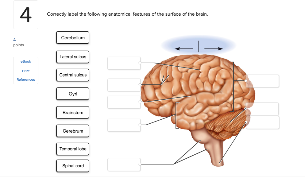
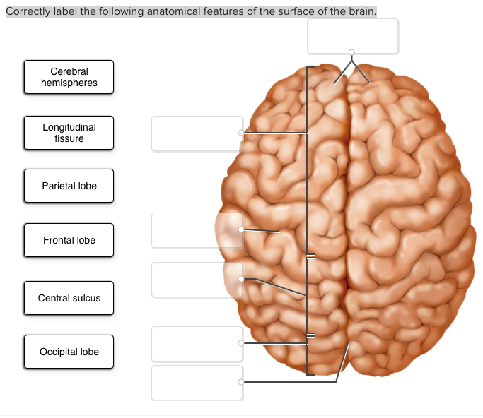
40 correctly label the following anatomical features of the surface of the brain.
AP1 Lab Manual_Answers - Anatomy and Physiology Lab Manual ... - StuDocu Anatomy and Physiology 3 (BIO255) Medical Terminology (HCM205) Managing Organizations and Leading People (C200 Task 1) World History (HY 390) Med Surg Clinical Lab Ii (NURS 636) Introduction To Computer And Information Security (ITO 310) Social Psychology (PSYC 215) Newest. Marketing Management (D174) Professional Application in Service ... F21F0CF9-8C36-45E3-A106-ED46F08D7C9F.jpeg - correctly label... View Homework Help - F21F0CF9-8C36-45E3-A106-ED46F08D7C9F.jpeg from EDU 46 at Bunker Hill Community College. correctly label the following anatomical features of the surface of the brain ." Spinal. Study Resources. Main Menu; by School; by Literature Title; by Subject; by Study Guides; Textbook Solutions Expert Tutors Earn.
PDF BIO 113 LAB 1. Anatomical Terminology, Positions, Planes, and Sections ... Surface Anatomy . Body surfaces provide a number of visible landmarks that can be used to study the body. Several of these are described on the following pages. Locating Body Landmarks . Anterior Body Landmarks . Identify and use anatomical terms to correctly label the following regions on Figure 1:

Correctly label the following anatomical features of the surface of the brain.
AFNI program: afni_proc.py - National Institutes of Health Feb 02, 2016 · DSET : name of input dataset, changed to copy_af_LABEL A default anatomical follower (in the case of skull stripping) is the original anat. That is to get a warped version that still has a skull, for quality control. See also -anat_follower_ROI, anat_follower_erode. -anat_follower_erode LABEL LABEL ...: erode masks for given labels Correctly label the following anatomical features of the surface of the ... Correctly label the following anatomical features of the surface of the brain. - Brainly.com. cliffbrown9056. 02/21/2022. Health. Solved 4 Correctly label the following anatomical features - Chegg Anatomy and Physiology questions and answers; 4 Correctly label the following anatomical features of the surface of the brain. Cerebellum 4 points Lateral sulcus eBook Print References Central sulcus Gyri Brainstem Cerebrum Temporal lobe Spinal cord ; Question: 4 Correctly label the following anatomical features of the surface of the brain ...
Correctly label the following anatomical features of the surface of the brain.. Brain Structure And Function | Brain Injury | British Columbia The Cerebellum - The cerebellum, or "little brain", is similar to the cerebrum with its two hemispheres and highly folded surface. It is associated with regulation and coordination of movement, posture, balance and cardiac, respiratory and vasomotor centers. Brain Stem - The brain stem is located beneath the limbic system. Anatomical Position and Directional Terms | Anatomy and Physiology The Anatomical Position First, let's talk about the anatomical position. The anatomical position is a standing position, with the head facing forward and the arms to the side. The palms are facing forward with the fingers extended, and the thumbs are pointing away from the body. The feet are spaced slightly apart with the toes pointing forward. Success Essays - Assisting students with assignments online Get 24⁄7 customer support help when you place a homework help service order with us. We will guide you on how to place your essay help, proofreading and editing your draft – fixing the grammar, spelling, or formatting of your paper easily and cheaply. Answered: ctly label the following veins of the… | bartleby ctly label the following veins of the thorax. Posterior ercostal veins Receives blood from the brain ernal jugular v. Subclavian v. Supreme intercostal v. Azygos v. Brachiocephalic V. Hemiazygos v. Question Transcribed Image Text: Special Circuits Saved Не Correctly label the following veins of the thorax.
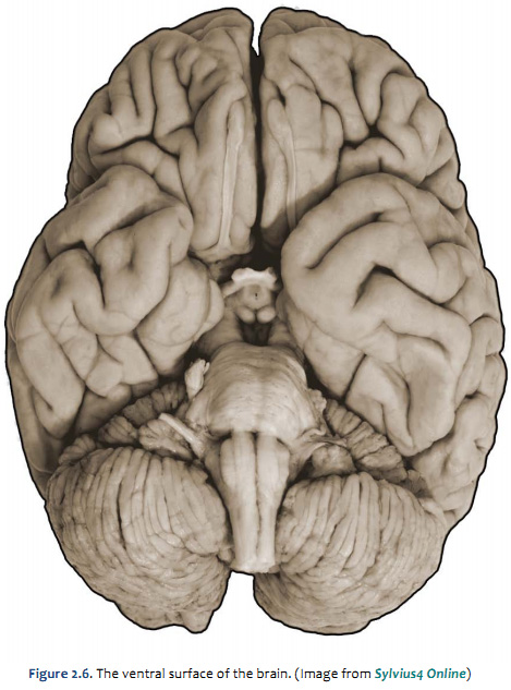
Achiever Papers - We help students improve their academic ... We offer assignment help in more than 80 courses. We are also able to handle any complex paper in any course as we have employed professional writers who are specialized in different fields of study. From their experience, they are able to work on the most difficult assignments. The following are some of the course we offer assignment help in ... 2,832 Labeled Brain Anatomy Images, Stock Photos & Vectors - Shutterstock Find Labeled brain anatomy stock images in HD and millions of other royalty-free stock photos, illustrations and vectors in the Shutterstock collection. Thousands of new, high-quality pictures added every day. Duke Neurosciences - Lab 2: Spinal Cord & Brainstem: Surface and ... Challenge 3.1—internal anatomy of the spinal cord. With reference to Figure 2.6, 2.7, and 2.8 and the chart below, carefully inspect the internal features of the spinal cord that are present in each segment, as well as those that are different (or present in only in one segment). To complete this challenge, spend some time browsing the spinal cord sections in Sylvius4, and find each of the ... Brain - Human Brain Diagrams and Detailed Information - Innerbody The forebrain (or prosencephalon) is made up of our incredible cerebrum, thalamus, hypothalamus and pineal gland among other features. Neuroanatomists call the cerebral area the telencephalon and use the term diencephalon (or interbrain) to refer to the area where our thalamus, hypothalamus and pineal gland reside.
Cross sectional anatomy | Kenhub Cross-sections are two-dimensional, axial views of gross anatomical structures seen in transverse planes. They are obtained by taking imaginary slices perpendicular to the main axis of organs, vessels, nerves, bones, soft tissue, or even the entire human body. Cross-sections provide the perception of 'depth', creating three-dimensional ... Main Parts of the Human Brain and Subdivisions of Human Brain Parts Thalamus, epithalamus, subthalamus and hypothalamus are the four sub-divisions. Here Hypothalamus is one of the parts of the human brain that initiates, coordinates, maintains and assists in the successful accomplishment of a number of visceral activities with the help of its hormonal secretions. Moreover, the sensory Optic Nerve, coming from ... 1.6 Anatomical Terminology - Anatomy and Physiology 2e - OpenStax Anterior (or ventral) Describes the front or direction toward the front of the body. The toes are anterior to the foot. Posterior (or dorsal) Describes the back or direction toward the back of the body. The popliteus is posterior to the patella. Superior (or cranial) describes a position above or higher than another part of the body proper. The Cerebellum - Structure - Position - TeachMeAnatomy The cerebellum, which stands for "little brain", is a structure of the central nervous system. It has an important role in motor control, with cerebellar dysfunction often presenting with motor signs. In particular, it is active in the coordination, precision and timing of movements, as well as in motor learning.
Free Science Flashcards about ANP1040 Exam 4 - StudyStack ANP1040 Exam 4. Correctly label the following anatomical features of a neuron. Correctly label the structures, areas, and concentrations associated with a cell's electrical charge difference across its membrane. ___ division carries signals to the smooth muscle in the large intestine.
Correctly Label the Following Anatomical Features of the Eye Correctly Label the Following Anatomical Features of the Eye In this article, you will learn the anatomical features of the eye, including the cornea. The optic nerve is the part of the eye that sends electrical signals from the eye to the brain. The eyeball is a sphere, containing three primary components: the retina, the pupil, and the lens.
Brain & CN Worksheet Flashcards | Quizlet Correctly label the following functional regions of the cerebral cortex. Consider a situation where a stroke or mechanical trauma has occurred resulting in damage to one of the areas of the brain indicated in the image. Drag each label into the proper location in order to identify the area that would most likely have been affected.
Anatomy of the Brain: Structures and Their Function - ThoughtCo The midbrain or mesencephalon, is the portion of the brainstem that connects the hindbrain and the forebrain. This region of the brain is involved in auditory and visual responses as well as motor function. The hindbrain extends from the spinal cord and is composed of the metencephalon and myelencephalon.
Anatomy and Physiology Questions and Answers - Study.com The study of the relationships of the body's structures by examining cross sections of tissues or organs is called [ {Blank}] anatomy. A) gross B) surface C) systemic D) regional E) sectional. View Answer. The study of the anatomical organization of specific areas of the body is called [ {Blank}] anatomy.
What are the three parts of the integumentary system. - Brainly.com Correctly label the following anatomical features of the surface of the brain. ... If someone faints, you should do all of the following except _____. Question 8 options: tighten the person's clothing. Leave the person lying dow … n and check the airway to make sure it is clear. Call for help if he or she does not regain consciousness in ...
Human Brain - Structure, Diagram, Parts Of Human Brain - BYJUS The human brain controls nearly every aspect of the human body ranging from physiological functions to cognitive abilities. It functions by receiving and sending signals via neurons to different parts of the body. The human brain, just like most other mammals, has the same basic structure, but it is better developed than any other mammalian brain.
Diagram of the Brain and its Functions - Bodytomy Functions. The frontal lobe is involved with the main executive functions of the brain, which include: Judgment, that is, the ability to recognize future consequences resulting from ongoing actions. This activity mostly occurs in the pre-frontal area. Analytical and critical reasoning.
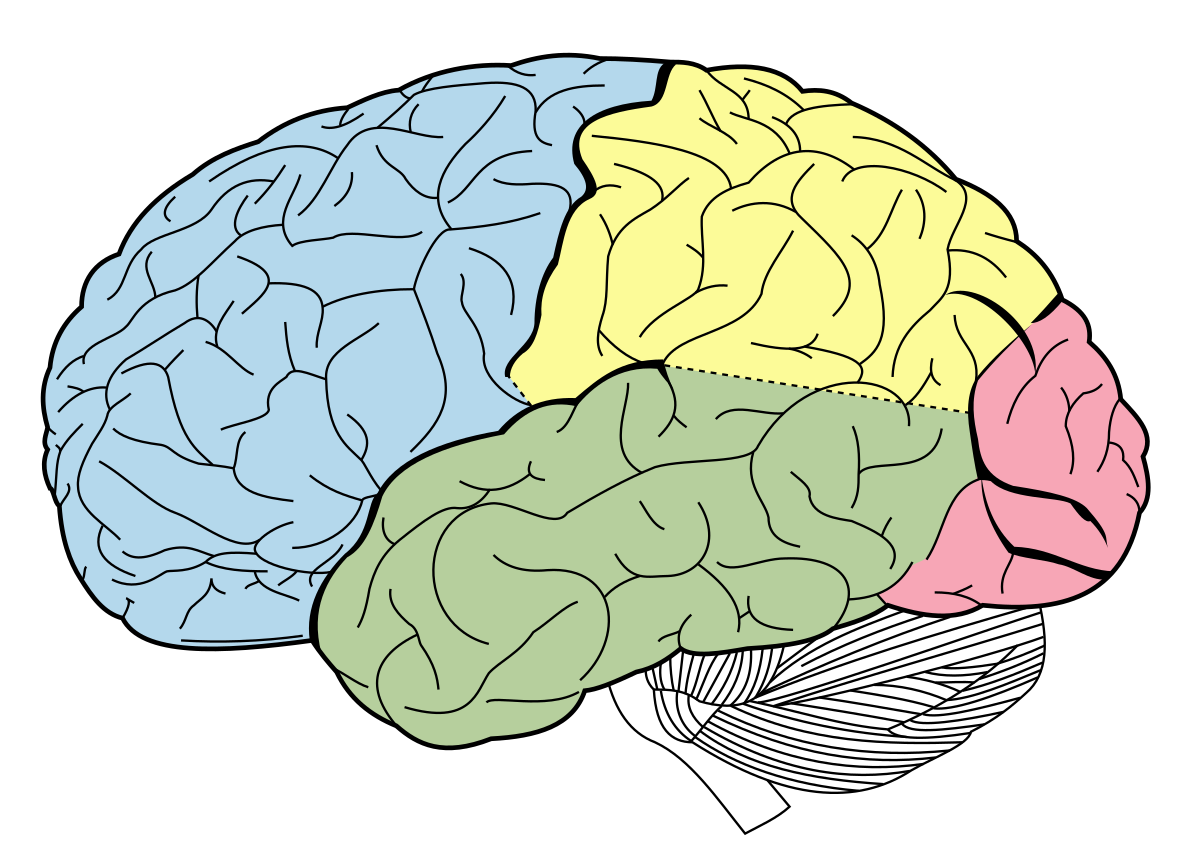
Lateral view of the brain: Anatomy and functions | Kenhub The lateral view of the brain shows the three major parts of the brain: cerebrum, cerebellum and brainstem . A lateral view of the cerebrum is the best perspective to appreciate the lobes of the hemispheres. Each hemisphere is conventionally divided into six lobes, but only four of them are visible from this lateral perspective.
8B4C53F7-CF59-48CF-BBD3-002197386B88.jpeg - Correctly label... View Homework Help - 8B4C53F7-CF59-48CF-BBD3-002197386B88.jpeg from BIO 203 at Bunker Hill Community College. Correctly label the following anatomical features of the surface of the brain. Cerebral
Assignment Essays - Best Custom Writing Services Get 24⁄7 customer support help when you place a homework help service order with us. We will guide you on how to place your essay help, proofreading and editing your draft – fixing the grammar, spelling, or formatting of your paper easily and cheaply.
Course Help Online - Have your academic paper written by a ... We offer assignment help in more than 80 courses. We are also able to handle any complex paper in any course as we have employed professional writers who are specialized in different fields of study. From their experience, they are able to work on the most difficult assignments. The following are some of the course we offer assignment help in ...
Chapter 13 QS Anatomy (Brain and Cranial Nerves) - Quizlet Controls muscular movement at the subconscious level=Putamen Correctly label the following anatomical features of the cerebellum. primary fissure, vermis, anterior lobe, posterior lobe, folia, cerebellar hemisphere Label the components of the cerebral nuclei. Claustrum, caudate nucleus, globus pallidus, putamen, amygdaloid body
PDF Brain Anatomy - Wou BI 335 - Advanced Human Anatomy and Physiology Western Oregon University Figure 4: Mid-sagittal section of brain showing diencephalon (includes corpus callosum, fornix, and anterior commissure) Marieb & Hoehn (Human Anatomy and Physiology, 9th ed.) - Figure 12.10 Exercise 2: Utilize the model of the human brain to locate the following structures / landmarks for the
Correctly Label the Following Anatomical Parts of Osseous Tissue. A typical long bone showing gross anatomical features. The wider section at each end of the bone is called the epiphysis (plural = epiphyses), which is filled internally with spongy bone, another type of osseous tissue. Red bone marrow fills the spaces between the spongy bone in some long bones. Each epiphysis meets the diaphysis at the metaphysis.
Solved correctly label the following anatomical features of - Chegg correctly label the following anatomical features of the surface of the brain. Show transcribed image text Expert Answer 100% (17 ratings) 1. Postcentral gyrus - Is on the lateral surface of parietal lobe. It lies parallel to the motor strip and is between the central sulcus and post central sulcus. 2.
Chapter 14 Worksheet Flashcards | Quizlet Study with Quizlet and memorize flashcards containing terms like Correctly label the following anatomical features of the surface of the brain., Correctly label the following meninges of the brain., Place a single word into each sentence to make it correct, then place each sentence into a logical paragraph order describing the flow of cerebrospinal fluid. and more.
Chapter 8/10 Flashcards | Quizlet Correctly label the anatomical features of the scapula. Label the structures of the bone using the hints provided. Correctly label the anatomical features of the femur and patella.
Solved 4 Correctly label the following anatomical features - Chegg Anatomy and Physiology questions and answers; 4 Correctly label the following anatomical features of the surface of the brain. Cerebellum 4 points Lateral sulcus eBook Print References Central sulcus Gyri Brainstem Cerebrum Temporal lobe Spinal cord ; Question: 4 Correctly label the following anatomical features of the surface of the brain ...
Correctly label the following anatomical features of the surface of the ... Correctly label the following anatomical features of the surface of the brain. - Brainly.com. cliffbrown9056. 02/21/2022. Health.
AFNI program: afni_proc.py - National Institutes of Health Feb 02, 2016 · DSET : name of input dataset, changed to copy_af_LABEL A default anatomical follower (in the case of skull stripping) is the original anat. That is to get a warped version that still has a skull, for quality control. See also -anat_follower_ROI, anat_follower_erode. -anat_follower_erode LABEL LABEL ...: erode masks for given labels






:watermark(/images/watermark_only_sm.png,0,0,0):watermark(/images/logo_url_sm.png,-10,-10,0):format(jpeg)/images/anatomy_term/superior-cerebral-veins/JROJBmhhQrzvz7nRxEWVQ_Superior_cerebral_veins_02__2_.png)





Post a Comment for "40 correctly label the following anatomical features of the surface of the brain."