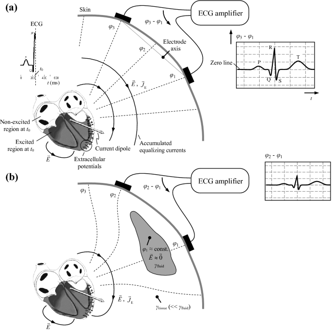
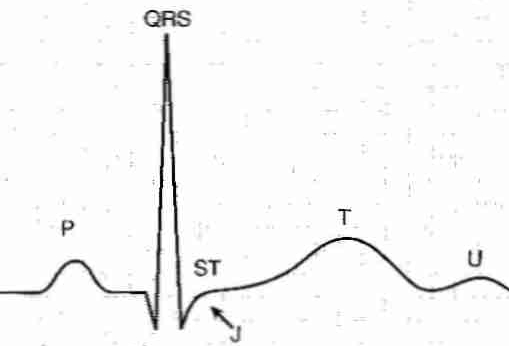
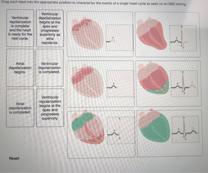
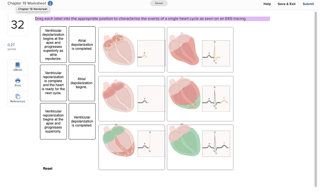
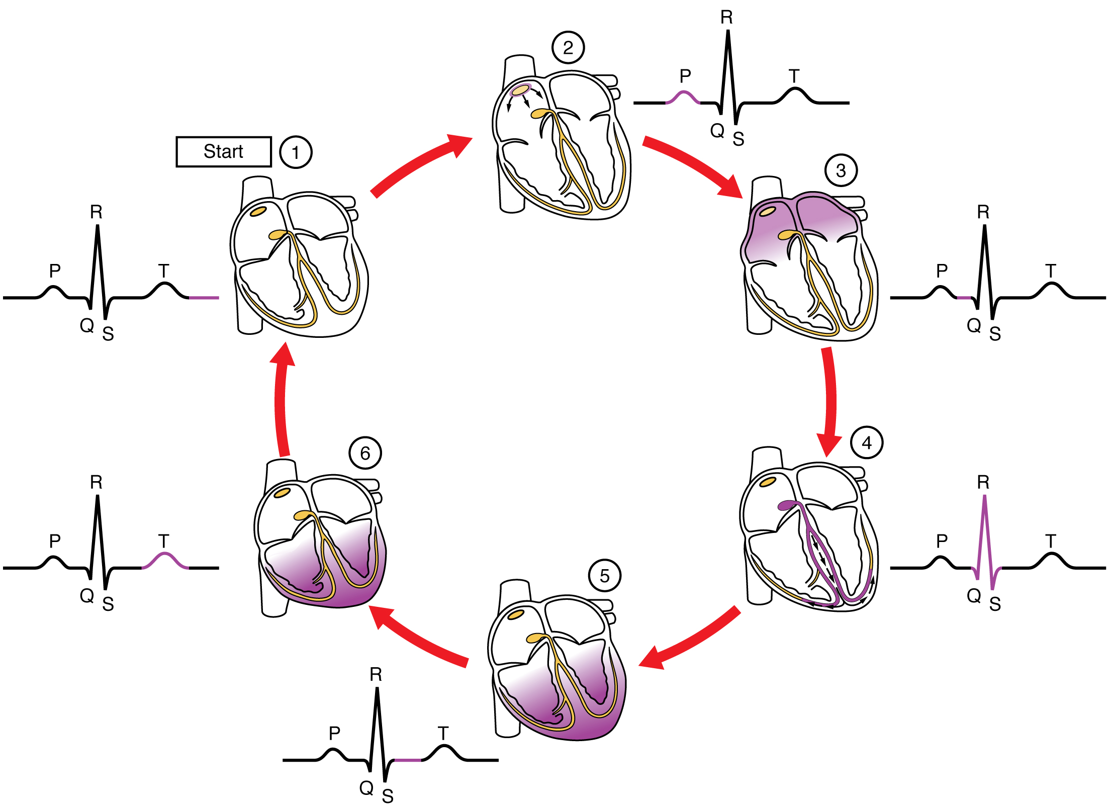
42 drag each label into the appropriate position to characterize the events of a single heart cycle as seen on an ecg tracing.
Solved Drag each label into the appropriate position to - Chegg Transcribed image text: Drag each label into the appropriate position to characterize the events of a single heart cycle as seen on an EKG tracing. Ventricular repolarization begins at the apex and progresses superiorly Ventricular repolarization is complete and the heart is ready for the next cycle Ventricular depolarization is completed Ventricular depolarization begins at the apex and ... Drag each label into the appropriate position to identify whether the ... Answer:• Apply a strip of masking tape, about 3 inches long, to the outside of each jar. Label each vial in writing on the masking tape with the marker. Write the following labels: hot water, cold water, RT water, vinegar, alcohol. • Add 1 cup of the appropriate liquid to each of the labeled jars.
quizlet.com › 571353173 › chapter-192021-flash-cardsChapter 19,20,21 Flashcards | Quizlet Study with Quizlet and memorize flashcards containing terms like Correctly label the pathway of blood flow through the heart, beginning with the right atrium., Indicate whether each structure is part of the systemic or pulmonary circuit., Drag each label into the appropriate position to characterize the events of a single heart cycle as seen on an ECG tracing. and more.
Drag each label into the appropriate position to characterize the events of a single heart cycle as seen on an ecg tracing.
Solved Saved Help Drag each label into the appropriate - Chegg This problem has been solved! See the answer See the answer See the answer done loading [Solved] Drag each label into the appropriate position to characterize ... Answered step-by-step Drag each label into the appropriate position to characterize the events of a single heart cycle as seen on an ECG tracing. Image transcription text Drag each label into the appropriate position to characterize the events of a single heart cycle as seen on an ECG tracing. SA node fires causing atrial depolarization in ECG interpretation: Characteristics of the normal ECG (P ... - ECG & ECHO The P-wave, PR interval and PR segment. ECG interpretation traditionally starts with an assessment of the P-wave. The P-wave reflects atrial depolarization (activation). The PR interval is the distance between the onset of the P-wave to the onset of the QRS complex. The PR interval is assessed in order to determine whether impulse conduction from the atria to the ventricles is normal.
Drag each label into the appropriate position to characterize the events of a single heart cycle as seen on an ecg tracing.. chaper 15 test 1 Flashcards | Quizlet Drag each label into the appropriate position to characterize the events of a single heart cycle as seen on an ECG tracing. In the cardiovascular system, what vessels are the site of nutrient, gas, and waste exchange? capillaries what is the deepest in the wall of the heart? Endocardium Complete the sentences describing the coverings of the heart. Solved Drag each label into the appropriate position to - Chegg question: drag each label into the appropriate position to characterize the events of a single heart cycle as seen on an ekg tracing ventricular ventricular depolarization repolarization begins at the is complete and the heart apex and progresses is ready for the superiorly as next cycle. atria repolanze atrial ventricular depolarization … › plans_allALEX | Alabama Learning Exchange m. Encode words with vowel y in the final position of one and two syllable words, distinguishing the difference between the long /imacr/ sound in one-syllable words and the long /emacr/ sound in two-syllable words, and words with vowel y in medial position, producing the short /ĭ/ sound for these words. quizlet.com › 574029087 › ch-19-circulatory-systemCh. 19 Circulatory System- heart Flashcards | Quizlet Correctly label the external anatomy of the anterior heart. Place the labels in order denoting the flow of blood through the pulmonary circuit beginning with the right atrium and ending in the left atrioventricular valve. The first and last structures are given. Right atrium 1. tricuspid valve 2. right ventricle 3. pulmonary valve
Ch. 20 Assignments.docx - Ch. 20 Practice - Lecture 1. Drag each label ... This preview shows page 1 - 4 out of 30 pages. View full document Ch. 20 Practice - Lecture 1. Drag each label into the appropriate position to identify whether the characteristic is indicative of arteries or veins. a. a. 2. Indicate whether the given condition would increase or decrease blood flow with all other factors being equal. a. a. 3. Chapter 15 Cardiovascular Practice Flashcards | Quizlet Complete each sentence, and then place them in the correct order to describe blood flow through the heart, beginning with blood entering the right side of the heart. Beginning with the return from the systemic circulation, blood enters the right atrium. Blood then travels through the tricuspid valve and into the right ventricle. quizlet.com › 184651599 › heart-lecture-flash-cardsHeart Lecture Flashcards | Quizlet Correctly label the following external anatomy of the posterior heart. Drag each label into the appropriate position to characterize the events of a single heart cycle as seen on an EKG tracing. Correctly label the pathway of blood flow through the heart, beginning with the right atrium. quizlet.com › 630625176 › chapter-19-the-heart-flashChapter 19: The Heart Flashcards | Quizlet -left heart—body—right heart supplies blood to all organs of the body Heart Location In the thoracic cavity, between the lungs in the mediastinum Size, Shape and Position of the Heart •Located in thoracic cavity -specifically in the mediastinum •area between lungs -superior to diaphragm -posterior to sternum
Solved Drag each label into the appropriate position to - Chegg Expert Answer. Parts of Normal ECG: 1. P wave: It denotes atrial depolarization. 2. QRS Complex: This is caused by ventricular depolarization. 3. Q wave: If the first wave of …. View the full answer. Transcribed image text: Drag each label into the appropriate position to identify the waves of a normal ECG Alpha wave 0 QRS complex ST segment ... Solved Drag each label into the appropriate position to - Chegg transcribed image text: chapter 19 worksheet g seved help save & exit submit chapter 19 worksheet drag each label into the appropriate position to characterize the events of a single heart cycle as seen on an ekg tracing 2 ventricular begins at the atrial apex and 0.27 points superiorly ass completed repolarize ventricular repolarization and the … quizlet.com › 259771705 › ch-15-cardiovascular-flashCh. 15 Cardiovascular Flashcards | Quizlet T wave Label the waves, or deflections, seen in the normal ECG pattern. Indicate whether each structure is part of the systemic circuit or the pulmonary circuit. The heart is located in the thoracic cavity The heart is situated between the _______ to either side, in front of the _____________________, and behind the __________________. Labeling a Normal ECG Drag each label into the appropriate position to ... 41) Labeling a Normal ECG. Drag each label into the appropriate position to characterize the events of a single heart cycle as seen on an ECG tracing. 42) If a person's heart is pumping 5000 mL of blood in one minute and the heart rate is 50 beats per minute, what is the cardiac output?
› createJoin LiveJournal Password requirements: 6 to 30 characters long; ASCII characters only (characters found on a standard US keyboard); must contain at least 4 different symbols;
Solved Drag each label into the appropriate position to - Chegg Drag each label into the appropriate position to characterize the events of a single heart cycle as seen on an ECG tracing. Show transcribed image text Expert Answer 100% (24 ratings) Figure 1 SA node fires causing atrial depolarization in right atrium Figure 5 ventricular de … View the full answer
Module 3.1.docx - 1. Describe in your own words what each... The ST segment is the flat line on the ECG; end of the S wave and beginning of the T wave. This segment is when ventricular depolarization and repolarization occurs. Mechanisms that may cause this to change may be due to different infections, such as Pericarditis or other abnormalities.
Ch 20 - Edited Cardiac Cycle, Part 2.pdf - Course Hero Cardiac cycle is complex and any points can be chosen as the beginningwhen heart is filled with blood Lubb and Dupp 140 Activity 2 Cardiovascular Problems 1. Atrial fibrillation is the rapid uncoordinated contraction of the atrial myocardium. In the space below, draw an ECG trace that would be characteristic of a atrial fibrillation. 2.
ECG interpretation: Characteristics of the normal ECG (P ... - ECG & ECHO The P-wave, PR interval and PR segment. ECG interpretation traditionally starts with an assessment of the P-wave. The P-wave reflects atrial depolarization (activation). The PR interval is the distance between the onset of the P-wave to the onset of the QRS complex. The PR interval is assessed in order to determine whether impulse conduction from the atria to the ventricles is normal.
[Solved] Drag each label into the appropriate position to characterize ... Answered step-by-step Drag each label into the appropriate position to characterize the events of a single heart cycle as seen on an ECG tracing. Image transcription text Drag each label into the appropriate position to characterize the events of a single heart cycle as seen on an ECG tracing. SA node fires causing atrial depolarization in
Solved Saved Help Drag each label into the appropriate - Chegg This problem has been solved! See the answer See the answer See the answer done loading















Post a Comment for "42 drag each label into the appropriate position to characterize the events of a single heart cycle as seen on an ecg tracing."